Labeled Muscles Of The Body Anterior View : Muscle Diagram German Text Male Body Muscle Chart German Labeling Most Important Muscles Of The Human Body Colored Canstock - Mikael häggström via wikimedia commons.. The cells of cardiac muscle tissue are striated—that is, they appear to have light and dark stripes when viewed under a light microscope. Learn about muscles anterior view superficial with free interactive flashcards. The muscles of the shoulder girdle are underdeveloped. Labeled muscles of the human body chart, anterior view, 3d rendering. Non small cell cancer of the lung.
The muscles of the anterior leg are located within the anterior compartment of the leg. Anterior view of superficial muscles of the body. Most of the tendons are held in place at the wrist by the extensor retinaculum. The style of citing shown here is from. The tibialis anterior muscle is located alongside the lateral surface of the tibia.
Naming Skeletal Muscles Anatomy And Physiology I from s3-us-west-2.amazonaws.com Superficial fascia of anterior abdominal wall: Anterior view.the superior nasal conchae are located posteriorly and are therefore not visible in the anterior view. What is the muscle labeled #1. Superficial+muscles+of+the+body+model+images | muscles of the human body (superficial short video of the anterior thigh muscles of the lower extremityidentifies:sartoriusquadriceps femoris the muscles of the leg anatomy chart shows in every possible view the way that the muscles and. The cells of cardiac muscle tissue are striated—that is, they appear to have light and dark stripes when viewed under a light microscope. There are four muscles in the anterior compartment of the leg: The sartorius is definitely labeled wrong. Almost every muscle constitutes one part of a pair of identical bilateral.
Superficial+muscles+of+the+body+model+images | muscles of the human body (superficial short video of the anterior thigh muscles of the lower extremityidentifies:sartoriusquadriceps femoris the muscles of the leg anatomy chart shows in every possible view the way that the muscles and. Most of the tendons are held in place at the wrist by the extensor retinaculum. Posterior compartment muscles of the forearm. The style of citing shown here is from. When observed macroscopically, this is seen as the because of that, contraction of these muscles will lead to a shortening of the muscle's body and cause the dorsum of the foot to be pulled towards the leg. Muscle attached to the fibula enabling the foot to extend and to draw away from the median axis of the body; When you research information you must cite the reference. Posteriorly, the vertebral line follows the spinal processes of the vertebra. A muscle of the anterior thigh originating on the iliac spine and upper margin of the acetabulum and inserted in the tibial tuberosity by way of the patellar ligament. This is an online quiz called muscles of the body anterior view. This is a table of skeletal muscles of the human anatomy. Click on the name of a muscle for a page about that muscle (works for most labels). Muscle that allows the big toe to extend and reinforces the action of the long extensor (extension of certain toes).
Muscle that allows the big toe to extend and reinforces the action of the long extensor (extension of certain toes). The anterior and middle scalene muscles, which also are located at the sides of the neck, act ipsilaterally to rotate. Produce wrist and/or finger flexion. The tibialis anterior muscle is located alongside the lateral surface of the tibia. Anterior view.the superior nasal conchae are located posteriorly and are therefore not visible in the anterior view.
Muscles Of The Neck And Torso Classic Human Anatomy In Motion The Artist S Guide To The Dynamics Of Figure Drawing from doctorlib.info Their main function is but muscle is also the dominant tissue in the heart and in the walls of other hollow organs of the body. When observed macroscopically, this is seen as the because of that, contraction of these muscles will lead to a shortening of the muscle's body and cause the dorsum of the foot to be pulled towards the leg. Anterior view.the superior nasal conchae are located posteriorly and are therefore not visible in the anterior view. Learn about muscles anterior view superficial with free interactive flashcards. The tibialis anterior muscle is located alongside the lateral surface of the tibia. The sartorius is definitely labeled wrong. Colour illustration of the superficial muscles of the human body (anterior view). It's pointing to a lower spot of the rectus femoris.
Citing for websites is different from citing from books, magazines and periodicals.
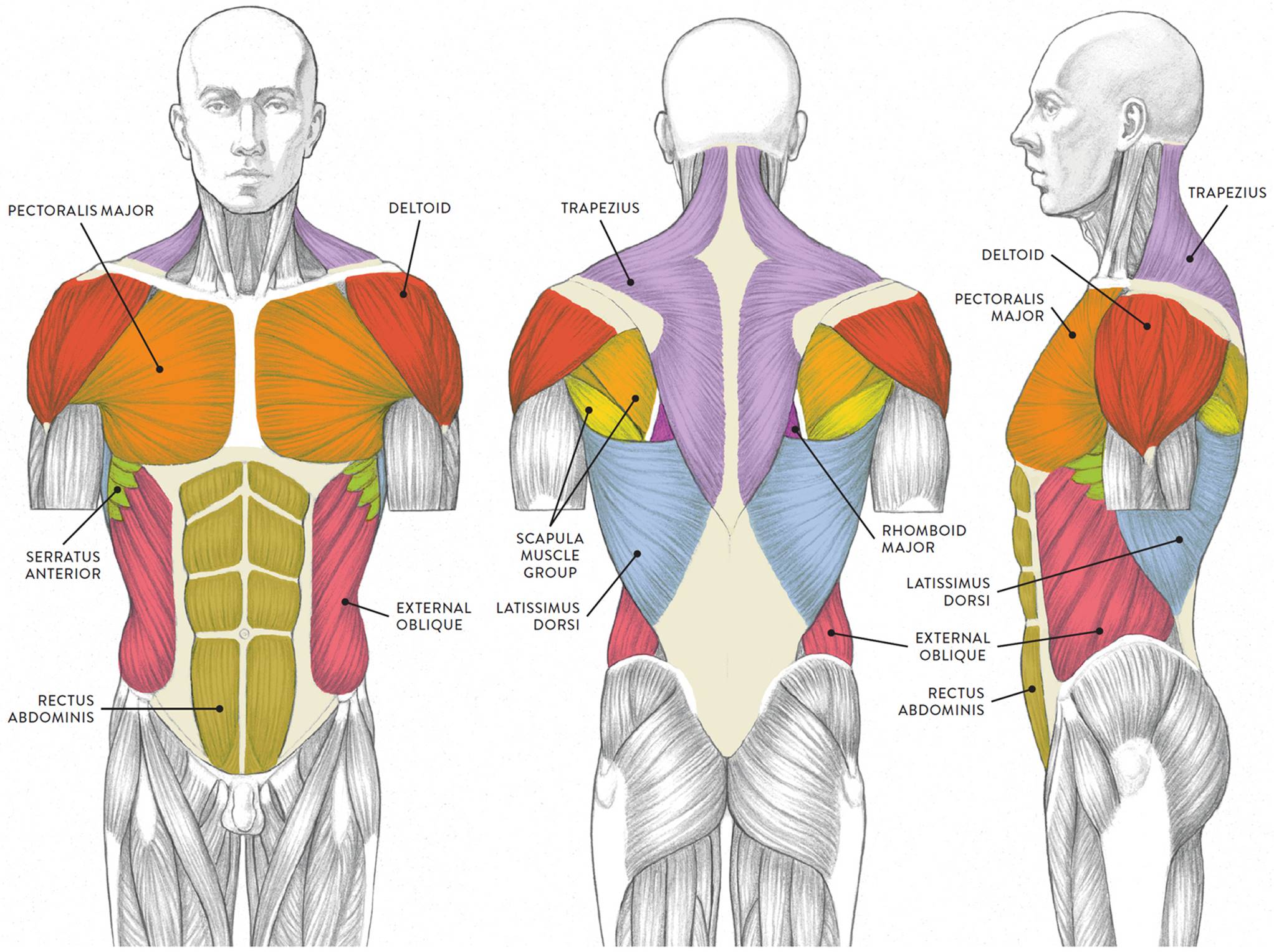
Frontalis, sartorius, pectoralis major, deltoid, thenar, biceps, rectus abdominis, serratus anterior, vastus lateralis, vastus medialis, rectus femorus, tibialis anterior, external obliques, brachioradialis, gastrocnemius, trapezius. Citing for websites is different from citing from books, magazines and periodicals. It also supports the plantar arch. The cells of cardiac muscle tissue are striated—that is, they appear to have light and dark stripes when viewed under a light microscope. Anterior muscles in the body. Arm anterior muscles labeled 3d illustration. Tibialis anterior muscle is a deep muscle of the leg arising from the tibia. Most of these originate from the lateral epicondyle. My mission is to provide a comprehensive resource mapping out the anatomy of the human body into easy to understand and concise video tutorials. Note that an interspace between two ribs is numbered by the rib above it. What is the muscle labeled #1. Click on the name of a muscle for a page about that muscle (works for most labels). The muscles of the shoulder girdle are underdeveloped.
Anterior muscles in the body. This first part covers the muscles of the anterior abdominal wall. Posteriorly, the vertebral line follows the spinal processes of the vertebra. Learn faster with these free muscle labeling diagrams. The rectus abdominis is a paired muscle running vertically on each side of the anterior.
Human Being Anatomy Muscles Anterior View Image Visual Dictionary from www.ikonet.com Human muscular system, labeling on the front view. Sternocleidomastoid trapezius serratus anterior latissimus dorsi pectoralis major pectoralis minor (deep muscle) rectus abdominus external. When you research information you must cite the reference. The anterior and middle scalene muscles, which also are located at the sides of the neck, act ipsilaterally to rotate. The thorax is longer than abdominal part of. Citing for websites is different from citing from books, magazines and periodicals. Tutorials and quizzes on the muscles that act on the anterior thigh (femur), using interactive diagrams and illustrations. Tibialis anterior, extensor digitorum longus, extensor hallucis longus and fibularis tertius.
Note that an interspace between two ribs is numbered by the rib above it.
Muscle that allows the big toe to extend and reinforces the action of the long extensor (extension of certain toes). There are around 650 skeletal muscles within the typical human body. Longus colli is a weak flexor the cervical spine and when contracting unilaterally it tilts and rotates the cervical spine to the ipsilateral side. The tibialis anterior muscle is located alongside the lateral surface of the tibia. Support and protect the abdominal viscera. Anterior view.the superior nasal conchae are located posteriorly and are therefore not visible in the anterior view. Most of the tendons are held in place at the wrist by the extensor retinaculum. This first part covers the muscles of the anterior abdominal wall. The sartorius is definitely labeled wrong. It's pointing to a lower spot of the rectus femoris. It also supports the plantar arch. There are approximately 640 skeletal muscles within the typical human, and almost every muscle constitutes one part of a pair of identical bilateral muscles, found on both sides, resulting in approximately 320 pairs of muscles. This muscle diagram is interactive:
Human muscular system, labeling on the front view anterior muscles of the body labeled. Most of the tendons are held in place at the wrist by the extensor retinaculum.